
Простой врожденный буллезный эпидермолиз (ВБЭ) включает в себя группу заболеваний, характеризующихся различной тяжестью течения, возможным поражением внутренних органов и различными исходами — от полного регресса высыпаний до смерти больного. Первоначальные клинические проявления простого ВБЭ не позволяют прогнозировать дальнейшее течение болезни, однако для этого могут быть использованы клинико-генетические корреляции. Проведен анализ литературы из баз данных PubMed и РИНЦ с целью характеристики клинико-генетических корреляций при простом ВБЭ. В результате проведенного анализа установлено, что наиболее тяжелое течение поражения кожи у больных простым ВБЭ ассоциируется с мутациями генов KRT5 и KRT14, изменяющими в соответствующих белках мотивы HIP (пептид инициации спирали) и HTP (пептид завершения спирали), а также спиральные участки кератинов 5 и 14. Отмечены также факторы, способные снижать надежность прогнозирования течения простого ВБЭ с использованием клинико-генетических корреляций. К ним относятся степень различий физико-химических свойств мутантной аминокислоты и аминокислоты дикого типа в случае миссенс-мутаций, а также возможное влияние вариантов других генов, способных участвовать в формировании клинической картины болезни. Выявление мутаций гена PLEC1 указывает на возможность развития с течением жизни у больного простым ВБЭ мышечной дистрофии, KLHL24 — кардиомиопатии, CD151 — нефропатии и глухоты. Таким образом, установлены клинико-генетические корреляции, которые могут быть использованы для прогнозирования течения простого ВБЭ, а также определены ограничения для их применения.
Предпросмотр статьи
Идентификаторы и классификаторы
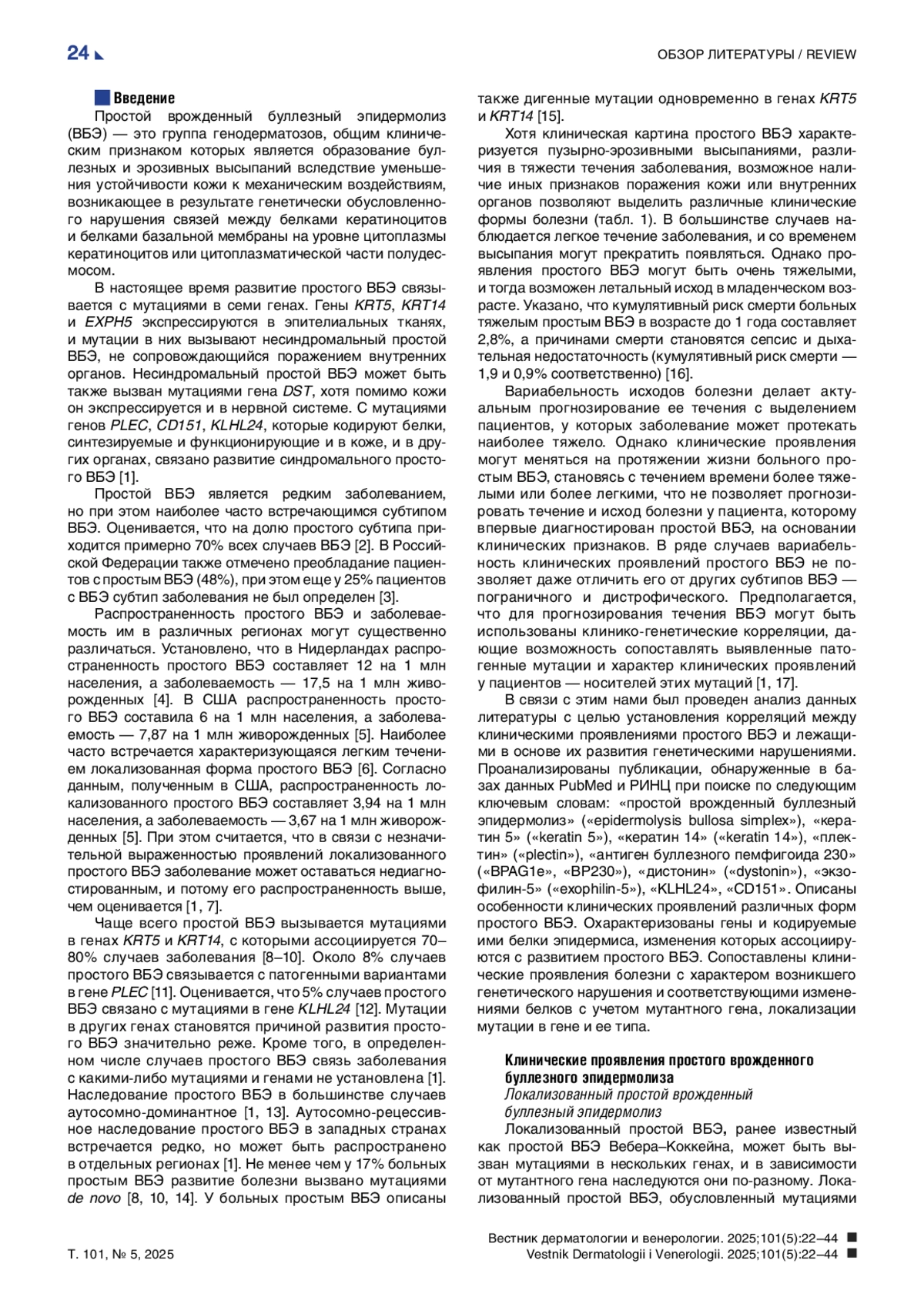
Простой врожденный буллезный эпидермолиз (ВБЭ) — это группа генодерматозов, общим клиническим признаком которых является образование буллезных и эрозивных высыпаний вследствие уменьшения устойчивости кожи к механическим воздействиям, возникающее в результате генетически обусловленного нарушения связей между белками кератиноцитов и белками базальной мембраны на уровне цитоплазмы кератиноцитов или цитоплазматической части полудесмосом
Если у вас возникли вопросы или появились предложения по содержанию статьи, пожалуйста, направляйте их в рамках данной темы.
Список литературы
1. Has C, Bauer JW, Bodemer C, Bolling MC, Bruckner-Tuderman L, Diem A, et al. Consensus reclassification of inherited epidermolysis bullosa and other disorders with skin fragility. Br J Dermatol. 2020;183(4):614-627. DOI: 10.1111/bjd.18921
2. Bardhan A, Bruckner-Tuderman L, Chapple IL, Fine JD, Harper N, Has C, et al. Epidermolysis bullosa. Nat Rev Dis Primers. 2020;6(1):78. DOI: 10.1038/s41572-020-0210-0
3. Кубанов А.А., Карамова А.Э., Чикин В.В., Богданова Е.В., Мончаковская Е.С. Эпидемиология и состояние оказания медицинской помощи больным врожденным буллезным эпидермолизом в Российской Федерации. Вестник РАМН. 2018;73(6):420-430. DOI: 10.15690/vramn980
4. Baardman R, Yenamandra VK, Duipmans JC, Pasmooij AMG, Jonkman MF, van den Akker PC, et al. Novel insights into the epidemiology of epidermolysis bullosa (EB) from the Dutch EB Registry: EB more common than previously assumed? J Eur Acad Dermatol Venereol. 2021;35(4):995-1006. DOI: 10.1111/jdv.17012
5. Fine JD. Epidemiology of Inherited Epidermolysis Bullosa Based on Incidence and Prevalence Estimates From the National Epidermolysis Bullosa Registry. JAMA Dermatol. 2016;152 (11):1231-1238. DOI: 10.1001/jamadermatol.2016.2473
6. Mariath LM, Santin JT, Schuler-Faccini L, Kiszewski AE. Inherited epidermolysis bullosa: update on the clinical and genetic aspects. An Bras Dermatol. 2020;95(5):551-569. DOI: 10.1016/j.abd.2020.05.001
7. Coulombe PA, Kerns ML, Fuchs E. Epidermolysis bullosa simplex: a paradigm for disorders of tissue fragility. J Clin Invest. 2009;119(7):1784-1793. DOI: 10.1172/JCI38177
8. Bolling MC, Lemmink HH, Jansen GH, Jonkman MF. Mutations in KRT5 and KRT14 cause epidermolysis bullosa simplex in 75% of the patients. Br J Dermatol. 2011;164(3):637-644. DOI: 10.1111/j.1365-2133.2010.10146.x
9. Rugg EL, Horn HM, Smith FJ, Wilson NJ, Hill AJ, Magee GJ, et al. Epidermolysis bullosa simplex in Scotland caused by a spectrum of keratin mutations. J Invest Dermatol. 2007;127(3):574-580. DOI: 10.1038/sj.jid.5700571
10. Wertheim-Tysarowska K, Ołdak M, Giza A, Kutkowska-Kaźmierczak A, Sota J, Przybylska D, et al. Novel sporadic and recurrent mutations in KRT5 and KRT14 genes in Polish epidermolysis bullosa simplex patients: further insights into epidemiology and genotype-phenotype correlation. J Appl Genet. 2016;57(2):175-181. DOI: 10.1007/s13353-015-0310-9
11. Bolling MC, Jongbloed JD, Boven LG, Diercks GF, Smith FJ, Irwin McLean WH, et al. Plectin mutations underlie epidermolysis bullosa simplex in 8% of patients. J Invest Dermatol. 2014;134(1):273-276. DOI: 10.1038/jid.2013.277
12. Has C, Fischer J. Inherited epidermolysis bullosa: New diagnostics and new clinical phenotypes. Exp Dermatol. 2019;28(10):1146-1152. DOI: 10.1111/exd.13668
13. So JY, Fulchand S, Wong CY, Li S, Nazaroff J, Gorell ES, et al. A global, cross-sectional survey of patient-reported outcomes, disease burden, and quality of life in epidermolysis bullosa simplex. Orphanet J Rare Dis. 2022;17(1):270. DOI: 10.1186/s13023-022-02433-3
14. Jerábková B, Marek J, Bucková H, Kopecková L, Veselý K, Valícková J, et al. Keratin mutations in patients with epidermolysis bullosa simplex: correlations between phenotype severity and disturbance of intermediate filament molecular structure. Br J Dermatol. 2010;162(5):1004-1013. DOI: 10.1111/j.1365-2133.2009.09626.x
15. Sathishkumar D, Orrin E, Terron-Kwiatkowski A, Browne F, Martinez AE, Mellerio JE, et al. The p.Glu477Lys mutation in keratin 5 is strongly associated with mortality in generalized severe epidermolysis bullosa simplex. J Invest Dermatol. 2016;136(3):719-721. DOI: 10.1016/j.jid.2015.11.024
16. Fine JD, Johnson LB, Weiner M, Suchindran C. Cause-specific risks of childhood death in inherited epidermolysis bullosa. J Pediatr. 2008;152(2):276-280. DOI: 10.1016/j.jpeds.2007.06.039
17. Uitto J, Atanasova VS, Jiang Q, South AP. Precision medicine for heritable skin diseases - The paradigm of epidermolysis bullosa. J Investig Dermatol Symp Proc. 2018;19(2):S74-S76. DOI: 10.1016/j.jisp.2018.09.004
18. Horn HM, Tidman MJ. The clinical spectrum of epidermolysis bullosa simplex. Br J Dermatol. 2000;142(3):468-472. DOI: 10.1046/j.1365-2133.2000.03358.x
19. Ganani D, Malovitski K, Sarig O, Gat A, Sprecher E, Samuelov L. Epidermolysis bullosa simplex due to bi-allelic DST mutations: Case series and review of the literature. Pediatr Dermatol. 2021;38(2):436-441. DOI: 10.1111/pde.14477
20. Karamatic Crew V, Burton N, Kagan A, Green CA, Levene C, Flinter F, et al. CD151, the first member of the tetraspanin (TM4) superfamily detected on erythrocytes, is essential for the correct assembly of human basement membranes in kidney and skin. Blood. 2004;104(8):2217-2223. DOI: 10.1182/blood-2004-04-1512
21. Vahidnezhad H, Youssefian L, Saeidian AH, Mahmoudi H, Touati A, Abiri M, et al. Recessive mutation in tetraspanin CD151 causes Kindler syndrome-like epidermolysis bullosa with multi-systemic manifestations including nephropathy. Matrix Biol. 2018;66:22-33. DOI: 10.1016/j.matbio.2017.11.003
22. Dunn C, Ambur A, Foss M, Nathoo R. Expanding the spectrum of epidermolysis bullosa simplex: Syndromic epidermolysis bullosa simplex with nephropathy and epilepsy secondary to CD151 tetraspanin defect-a case report and review of the literature. JAAD Case Rep. 2022;23:136-140. DOI: 10.1016/j.jdcr.2022.03.012
23. Liu L, Dopping-Hepenstal PJ, Lovell PA, Michael M, Horn H, Fong K, et al. Autosomal recessive epidermolysis bullosa simplex due to loss of BPAG1-e expression. J Invest Dermatol. 2012;132(3Pt 1):742-744. DOI: 10.1038/jid.2011.379
24. He Y, Leppert J, Steinke H, Has C. Homozygous nonsense mutation and additional deletion of an amino acid in BPAG1e causing mild localized epidermolysis bullosa simplex. Acta Derm Venereol. 2017;97(5):657-659. DOI: 10.2340/00015555-2618
25. Fine JD, Bruckner-Tuderman L, Eady RA, Bauer EA, Bauer JW, Has C, et al. Inherited epidermolysis bullosa: updated recommendations on diagnosis and classification. J Am Acad Dermatol. 2014;70(6):1103-1126. DOI: 10.1016/j.jaad.2014.01.903
26. Almokali K, Alshalawi H, Aldriwesh MG, Alotibi RS. Nephrotic syndrome: Pretibial epidermolysis bullosa in a patient with CD151 tetraspanin defect: A case report. Int J Health Sci (Qassim). 2024;18(1):35-40.
27. McGrath JA, Ishida-Yamamoto A, Tidman MJ, Heagerty AH, Schofield OM, Eady RA. Epidermolysis bullosa simplex (Dowling-Meara). A clinicopathological review. Br J Dermatol. 1992;126(5):421-430. DOI: 10.1111/j.1365-2133.1992.tb11813.x
28. Arin MJ. The molecular basis of human keratin disorders. Hum Genet. 2009;125(4):355-373. DOI: 10.1007/s00439-009-0646-5
29. Mithwani AA, Hashmi A, Adil S. Epidermolysis bullosa and congenital pyloric atresia. BMJ Case Rep. 2013;2013:bcr2013201207. DOI: 10.1136/bcr-2013-201207
30. Parelkar SV, Kapadnis SP, Sanghvi BV, Joshi PB, Mundada D, Shetty S, et al. Pyloric atresia - three cases and review of literature. Afr J Paediatr Surg. 2014;11(4):362-365. DOI: 10.4103/0189-6725.143178
31. Natsuga K. Plectin-related skin diseases. J Dermatol Sci. 2015;77(3):139-145. DOI: 10.1016/j.jdermsci.2014.11.005
32. Sawamura D, Goto M, Sakai K, Nakamura H, McMillan JR, Akiyama M, et al. Possible involvement of exon 31 alternative splicing in phenotype and severity of epidermolysis bullosa caused by mutations in PLEC1. J Invest Dermatol. 2007;127(6):1537-1540. DOI: 10.1038/sj.jid.5700707
33. Charlesworth A, Chiaverini C, Chevrant-Breton J, DelRio M, Diociaiuti A, Dupuis RP, et al. Epidermolysis bullosa simplex with PLEC mutations: new phenotypes and new mutations. Br J Dermatol. 2013;168(4):808-814. DOI: 10.1111/bjd.12202
34. Pfendner E, Uitto J. Plectin gene mutations can cause epidermolysis bullosa with pyloric atresia. J Invest Dermatol. 2005;124(1):111-115. DOI: 10.1111/j.0022-202X.2004.23564.x
35. Turcan I, Pasmooij AM, Gostyński A, van den Akker PC, Lemmink HH, Diercks GF, et al. Epidermolysis bullosa simplex caused by distal truncation of BPAG1-e: An intermediate generalized phenotype with prurigo papules. J Invest Dermatol. 2017;137(10):2227-2230. DOI: 10.1016/j.jid.2017.04.041
36. Diociaiuti A, Pisaneschi E, Rossi S, Condorelli AG, Carnevale C, Zambruno G, et al. Autosomal recessive epidermolysis bullosa simplex due to EXPH5 mutation: neonatal diagnosis of the first Italian case and literature review. J Eur Acad Dermatol Venereol. 2020;34(11):e694-e697. DOI: 10.1111/jdv.16372
37. McGrath JA, Stone KL, Begum R, Simpson MA, Dopping-Hepenstal PJ, Liu L, et al. Germline mutation in EXPH5 implicates the Rab27B effector protein Slac2-b in inherited skin fragility. Am J Hum Genet. 2012;91(6):1115-1121. DOI: 10.1016/j.ajhg.2012.10.012
38. Lee JY, Liu L, Hsu CK, Aristodemou S, Ozoemena L, Ogboli M, et al. Mutations in KLHL24 add to the molecular heterogeneity of epidermolysis bullosa simplex. J Invest Dermatol. 2017;137(6):1378-1380. DOI: 10.1016/j.jid.2017.01.004
39. Mariath LM, Santin JT, Frantz JA, Doriqui MJ, Schuler-Faccini L, Kiszewski AE. Genotype-phenotype correlations on epidermolysis bullosa with congenital absence of skin: A comprehensive review. Clin Genet. 2021;99(1):29-41. DOI: 10.1111/cge.13792
40. He Y, Maier K, Leppert J, Hausser I, Schwieger-Briel A, Weibel L, et al. Monoallelic mutations in the translation initiation codon of KLHL24 cause skin fragility. Am J Hum Genet. 2016;99(6):1395-1404. DOI: 10.1016/j.ajhg.2016.11.005
41. Lin Z, Li S, Feng C, Yang S, Wang H, Ma D, et al. Stabilizing mutations of KLHL24 ubiquitin ligase cause loss of keratin 14 and human skin fragility. Nat Genet. 2016;48(12):1508-1516. DOI: 10.1038/ng.3701
42. El Hachem M, Barresi S, Diociaiuti A, Boldrini R, Condorelli AG, Capoluongo E, et al. Phenotypic features of epidermolysis bullosa simplex due to KLHL24 mutations in 3 Italian cases. Acta Derm Venereol. 2019;99(2):238-239. DOI: 10.2340/00015555-3046
43. Yenamandra VK, van den Akker PC, Lemmink HH, Jan SZ, Diercks GFH, Vermeer M, et al. Cardiomyopathy in patients with epidermolysis bullosa simplex with mutations in KLHL24. Br J Dermatol. 2018;179(5):1181-1183. DOI: 10.1111/bjd.16797
44. Schwieger-Briel A, Fuentes I, Castiglia D, Barbato A, Greutmann M, Leppert J, et al. Epidermolysis bullosa simplex with KLHL24 mutations is associated with dilated cardiomyopathy. J Invest Dermatol. 2019;139(1):244-249. DOI: 10.1016/j.jid.2018.07.022
45. Alkhalifah A, Chiaverini C, Charlesworth A, Has C, Lacour JP. Burnlike scars: A sign suggestive of KLHL24-related epidermolysis bullosa simplex. Pediatr Dermatol. 2018;35(3):e193-e195. DOI: 10.1111/pde.13443
46. Bolling MC, Jonkman MF. KLHL24: Beyond skin fragility. J Invest Dermatol. 2019;139(1):22-24. DOI: 10.1016/j.jid.2018.08.010
47. Burke MA, Cook SA, Seidman JG, Seidman CE. Clinical and mechanistic insights into the genetics of cardiomyopathy. J Am Coll Cardiol. 2016;68(25):2871-2886. DOI: 10.1016/j.jacc.2016.08.079
48. Kyrova J, Kopeckova L, Buckova H, Mrazova L, Vesely K, Hermanova M, et al. Epidermolysis bullosa simplex with muscular dystrophy. Review of the literature and a case report. J Dermatol Case Rep. 2016;10(3):39-48. DOI: 10.3315/jdcr.2016.1231
49. Gache Y, Chavanas S, Lacour JP, Wiche G, Owaribe K, Meneguzzi G, et al. Defective expression of plectin/HD1 in epidermolysis bullosa simplex with muscular dystrophy. J Clin Invest. 1996;97(10):2289-2298. DOI: 10.1172/JCI118671
50. Fischer T, Gedde-Dahl T Jr. Epidermolysis bullosa simplex and mottled pigmentation: a new dominant syndrome. I. Clinical and histological features. Clin Genet. 1979;15(3):228-238. DOI: 10.1111/j.1399-0004.1979.tb00972.x
51. Westerhof W, Dingemans KP. Generalized mottled pigmentation with postnatal skin blistering in three generations. J Am Acad Dermatol. 2004;50(5Suppl):S65-69. DOI: 10.1016/j.jaad.2003.07.015
52. Bruckner-Tuderman L, Vogel A, Rüegger S, Odermatt B, Tönz O, Schnyder UW. Epidermolysis bullosa simplex with mottled pigmentation. J Am Acad Dermatol. 1989;21(2Pt 2):425-432. DOI: 10.1016/s0190-9622(89)80052-0
53. Echeverría-García B, Vicente A, Hernández Á, Mascaró JM, Colmenero I, Terrón A, et al. Epidermolysis bullosa simplex with mottled pigmentation: a family report and review. Pediatr Dermatol. 2013;30(6):e125-31. DOI: 10.1111/j.1525-1470.2012.01748.x
54. Combemale P, Kanitakis J. Epidermolysis bullosa simplex with mottled pigmentation. Case report and review of the literature. Dermatology. 1994;189(2):173-178. DOI: 10.1159/000246826
55. Coleman R, Harper JI, Lake BD. Epidermolysis bullosa simplex with mottled pigmentation. Br J Dermatol. 1993;128(6):679-685. DOI: 10.1111/j.1365-2133.1993.tb00265.x
56. Horiguchi Y, Sawamura D, Mori R, Nakamura H, Takahashi K, Shimizu H. Clinical heterogeneity of 1649delG mutation in the tail domain of keratin 5: a Japanese family with epidermolysis bullosa simplex with mottled pigmentation. J Invest Dermatol. 2005;125(1):83-85. DOI: 10.1111/j.0022-202X.2005.23790.x
57. Andres C, Chen W, Hofmann H, Ring J, Schnopp C. Epidermolysis bullosa simplex with mottled pigmentation: a case report. Int J Dermatol. 2009;48(7):753-754. DOI: 10.1111/j.1365-4632.2009.03846.x
58. Harel A, Bergman R, Indelman M, Sprecher E. Epidermolysis bullosa simplex with mottled pigmentation resulting from a recurrent mutation in KRT14. J Invest Dermatol. 2006;126(7):1654-1657. DOI: 10.1038/sj.jid.5700296
59. Uttam J, Hutton E, Coulombe PA, Anton-Lamprecht I, Yu QC, Gedde-Dahl T Jr, et al. The genetic basis of epidermolysis bullosa simplex with mottled pigmentation. Proc Natl Acad Sci U S A. 1996;93(17):9079-9084. DOI: 10.1073/pnas.93.17.9079
60. Irvine AD, Rugg EL, Lane EB, Hoare S, Peret C, Hughes AE, et al. Molecular confirmation of the unique phenotype of epidermolysis bullosa simplex with mottled pigmentation. Br J Dermatol. 2001;144(1):40-45. DOI: 10.1046/j.1365-2133.2001.03950.x
61. Hamada T, Ishii N, Kawano Y, Takahashi Y, Inoue M, Yasumoto S, et al. The P25L mutation in the KRT5 gene in a Japanese family with epidermolysis bullosa simplex with mottled pigmentation. Br J Dermatol. 2004;150(3):609-611. DOI: 10.1046/j.1365-2133.2004.05820.x
62. Pascucci M, Posteraro P, Pedicelli C, Provini A, Auricchio L, Paradisi M, et al. Epidermolysis bullosa simplex with mottled pigmentation due to de novo P25L mutation in keratin 5 in an Italian patient. Eur J Dermatol. 2006;16(6):620-622.
63. Irvine AD, McKenna KE, Jenkinson H, Hughes AE. A mutation in the V1 domain of keratin 5 causes epidermolysis bullosa simplex with mottled pigmentation. J Invest Dermatol. 1997;108(5):809-810. DOI: 10.1111/1523-1747.ep12292263
64. Tang HY, Du WD, Cui Y, Fan X, Quan C, Fang QY, et al. One novel and two recurrent mutations in the keratin 5 gene identified in Chinese patients with epidermolysis bullosa simplex. Clin Exp Dermatol. 2009;34(8):e957-961. DOI: 10.1111/j.1365-2230.2009.03703.x
65. Moog U, de Die-Smulders CE, Scheffer H, van der Vlies P, Henquet CJ, Jonkman MF. Epidermolysis bullosa simplex with mottled pigmentation: clinical aspects and confirmation of the P24L mutation in the KRT5 gene in further patients. Am J Med Genet. 1999;86(4):376-379. :4<376::aid-ajmg12>3.0.co;2-w. DOI: 10.1002/(sici)1096-8628(19991008)86
66. Yasukawa K, Sawamura D, Akiyama M, Motoda N, Shimizu H. Keratotic lesions in epidermolysis bullosa simplex with mottled pigmentation. J Am Acad Dermatol. 2005;52(1):172-173. DOI: 10.1016/j.jaad.2004.07.046
67. Shurman D, Losi-Sasaki J, Grimwood R, Kivirikko S, Tichy E, Uitto J, et al. Epidermolysis bullosa simplex with mottled pigmentation: mutation analysis in the first reported Hispanic pedigree with the largest single generation of affected individuals to date. Eur J Dermatol. 2006;16(2):132-135.
68. Kumagai Y, Umegaki-Arao N, Sasaki T, Nakamura Y, Takahashi H, Ashida A, et al. Distinct phenotype of epidermolysis bullosa simplex with infantile migratory circinate erythema due to frameshift mutations in the V2 domain of KRT5. J Eur Acad Dermatol Venereol. 2017;31(5):e241-e243. DOI: 10.1111/jdv.14005
69. Yalici-Armagan B, Kabacam S, Taskiran ZE, Gököz Ö, Utine GE, Ersoy-Evans S. A novel mutation of keratin 5 in epidermolysis bullosa simplex with migratory circinate erythema. Pediatr Dermatol. 2020;37(2):358-361. DOI: 10.1111/pde.14087
70. Lee SE, Choi JY, Kim SE, Kim SC. A novel deletion mutation in the 2B domain of KRT5 in epidermolysis bullosa simplex with childhood-onset migratory circinate erythema. Eur J Dermatol. 2018;28(1):123-125. DOI: 10.1684/ejd.2017.3190
71. Winter L, Wiche G. The many faces of plectin and plectinopathies: pathology and mechanisms. Acta Neuropathol. 2013;125(1):77-93. DOI: 10.1007/s00401-012-1026-0
72. Leung CL, Zheng M, Prater SM, Liem RK. The BPAG1 locus: Alternative splicing produces multiple isoforms with distinct cytoskeletal linker domains, including predominant isoforms in neurons and muscles. J Cell Biol. 2001;154(4):691-697. DOI: 10.1083/jcb.200012098
73. Walko G, Castañón MJ, Wiche G. Molecular architecture and function of the hemidesmosome. Cell Tissue Res. 2015;360(3):529-544. DOI: 10.1007/s00441-015-2216-6
74. Leung CL, Liem RK, Parry DA, Green KJ. The plakin family. J Cell Sci. 2001;114(Pt 19):3409-3410. DOI: 10.1242/jcs.114.19.3409
75. Koster J, Geerts D, Favre B, Borradori L, Sonnenberg A. Analysis of the interactions between BP180, BP230, plectin and the integrin alpha6beta4 important for hemidesmosome assembly. J Cell Sci. 2003;116(Pt 2):387-399. DOI: 10.1242/jcs.00241
76. Moll R, Divo M, Langbein L. The human keratins: biology and pathology. Histochem Cell Biol. 2008;129(6):705-733. DOI: 10.1007/s00418-008-0435-6
77. Chamcheu JC, Siddiqui IA, Syed DN, Adhami VM, Liovic M, Mukhtar H. Keratin gene mutations in disorders of human skin and its appendages. Arch Biochem Biophys. 2011;508(2):123-137. DOI: 10.1016/j.abb.2010.12.019
78. Herrmann H, Aebi U. Intermediate filaments: Structure and assembly. Cold Spring Harb Perspect Biol. 2016;8(11):a018242. DOI: 10.1101/cshperspect.a018242
79. Liovic M, Stojan J, Bowden PE, Gibbs D, Vahlquist A, Lane EB, et al. A novel keratin 5 mutation (K5V186L) in a family with EBS-K: a conservative substitution can lead to development of different disease phenotypes. J Invest Dermatol. 2001;116(6):964-969. DOI: 10.1046/j.1523-1747.2001.01334.x
80. Yasukawa K, Sawamura D, McMillan JR, Nakamura H, Shimizu H. Dominant and recessive compound heterozygous mutations in epidermolysis bullosa simplex demonstrate the role of the stutter region in keratin intermediate filament assembly. J Biol Chem. 2002;277(26):23670-23674. DOI: 10.1074/jbc.M200974200
81. Jankowski M, Wertheim-Tysarowska K, Jakubowski R, Sota J, Nowak W, Czajkowski R. Novel KRT14 mutation causing epidermolysis bullosa simplex with variable phenotype. Exp Dermatol. 2014;23(9):684-687. DOI: 10.1111/exd.12478
82. Leung CL, Green KJ, Liem RK. Plakins: a family of versatile cytolinker proteins. Trends Cell Biol. 2002;12(1):37-45. DOI: 10.1016/s0962-8924(01)02180-8
83. Eldirany SA, Lomakin IB, Ho M, Bunick CG. Recent insight into intermediate filament structure. Curr Opin Cell Biol. 2021;68:132-143. DOI: 10.1016/j.ceb.2020.10.001
84. Omary MB, Ku NO, Tao GZ, Toivola DM, Liao J. “Heads and tails” of intermediate filament phosphorylation: multiple sites and functional insights. Trends Biochem Sci. 2006;31(7):383-394. DOI: 10.1016/j.tibs.2006.05.008
85. Wu KC, Bryan JT, Morasso MI, Jang SI, Lee JH, Yang JM, et al. Coiled-coil trigger motifs in the 1B and 2B rod domain segments are required for the stability of keratin intermediate filaments. Mol Biol Cell. 2000;11(10):3539-3358. DOI: 10.1091/mbc.11.10.3539
86. Banerjee S, Wu Q, Yu P, Qi M, Li C. In silico analysis of all point mutations on the 2B domain of K5/K14 causing epidermolysis bullosa simplex: a genotype-phenotype correlation. Mol Biosyst. 2014;10(10):2567-2577. DOI: 10.1039/c4mb00138a
87. Lee CH, Kim MS, Chung BM, Leahy DJ, Coulombe PA. Structural basis for heteromeric assembly and perinuclear organization of keratin filaments. Nat Struct Mol Biol. 2012;19(7):707-715. DOI: 10.1038/nsmb.2330
88. Müller FB, Küster W, Wodecki K, Almeida H Jr, Bruckner-Tuderman L, Krieg T, et al. Novel and recurrent mutations in keratin KRT5 and KRT14 genes in epidermolysis bullosa simplex: implications for disease phenotype and keratin filament assembly. Hum Mutat. 2006;27(7):719-720. DOI: 10.1002/humu.9437
89. Fuchs E, Weber K. Intermediate filaments: structure, dynamics, function, and disease. Annu Rev Biochem. 1994;63:345-382. DOI: 10.1146/annurev.bi.63.070194.002021
90. Planko L, Böhse K, Höhfeld J, Betz RC, Hanneken S, Eigelshoven S, et al. Identification of a keratin-associated protein with a putative role in vesicle transport. Eur J Cell Biol. 2007;86(11-12):827-839. DOI: 10.1016/j.ejcb.2007.02.004
91. Coulombe PA, Lee CH. Defining keratin protein function in skin epithelia: epidermolysis bullosa simplex and its aftermath. J Invest Dermatol. 2012;132(3Pt 2):763-775. DOI: 10.1038/jid.2011.450
92. Has C, Schumann H, Leppert J, He Y, Hartmann B, Hausser I, et al. Monoallelic Large Intragenic KRT5 Deletions Account for Genetically Unsolved Cases of Epidermolysis Bullosa Simplex. J Invest Dermatol. 2017;137(10):2231-2234. DOI: 10.1016/j.jid.2017.05.016
93. Titeux M, Mazereeuw-Hautier J, Hadj-Rabia S, Prost C, Tonasso L, Fraitag S, et al. Three severe cases of EBS Dowling-Meara caused by missense and frameshift mutations in the keratin 14 gene. J Invest Dermatol. 2006;126(4):773-776. DOI: 10.1038/sj.jid.5700154
94. Arin MJ, Grimberg G, Schumann H, De Almeida H Jr, Chang YR, Tadini G, et al. Identification of novel and known KRT5 and KRT14 mutations in 53 patients with epidermolysis bullosa simplex: correlation between genotype and phenotype. Br J Dermatol. 2010;162(6):1365-1369. DOI: 10.1111/j.1365-2133.2010.09657.x
95. Szeverenyi I, Cassidy AJ, Chung CW, Lee BT, Common JE, Ogg SC, et al. The Human Intermediate Filament Database: comprehensive information on a gene family involved in many human diseases. Hum Mutat. 2008;29(3):351-360. DOI: 10.1002/humu.20652
96. Steinert PM. Structure, function, and dynamics of keratin intermediate filaments. J Invest Dermatol. 1993;100(6):729-734. DOI: 10.1111/1523-1747.ep12475665
97. Steinert PM, Marekov LN, Fraser RD, Parry DA. Keratin intermediate filament structure. Crosslinking studies yield quantitative information on molecular dimensions and mechanism of assembly. J Mol Biol. 1993;230(2):436-452. DOI: 10.1006/jmbi.1993.1161
98. Shinkuma S, Nishie W, Jacyk WK, Natsuga K, Ujiie H, Nakamura H, et al. A novel keratin 5 mutation in an African family with epidermolysis bullosa simplex indicates the importance of the amino acid located at the boundary site between the H1 and coil 1A domains. Acta Derm Venereol. 2013;93(5):585-587. DOI: 10.2340/00015555-1538
99. Ołdak M, Szczecińska W, Przybylska D, Maksym RB, Podgórska M, Woźniak K, et al. Gene dosage effect of p.Glu170Lys mutation in the KRT5 gene in a Polish family with epidermolysis bullosa simplex. J Dermatol Sci. 2011;61(1):64-67. DOI: 10.1016/j.jdermsci.2010.11.002
100. Cummins RE, Klingberg S, Wesley J, Rogers M, Zhao Y, Murrell DF. Keratin 14 point mutations at codon 119 of helix 1A resulting in different epidermolysis bullosa simplex phenotypes. J Invest Dermatol. 2001;117(5):1103-1107. DOI: 10.1046/j.0022-202x.2001.01508.x
101. Natsuga K, Nishie W, Smith BJ, Shinkuma S, Smith TA, Parry DA, et al. Consequences of two different amino-acid substitutions at the same codon in KRT14 indicate definitive roles of structural distortion in epidermolysis bullosa simplex pathogenesis. J Invest Dermatol. 2011;131(9):1869-1876. DOI: 10.1038/jid.2011.143
102. Lalor L, Titeux M, Palisson F, Fuentes I, Yubero MJ, Tasanen K, et al. Epidermolysis bullosa simplex-generalized severe type due to keratin 5 p.Glu477Lys mutation: Genotype-phenotype correlation and in silico modeling analysis. Pediatr Dermatol. 2019;36(1):132-138. DOI: 10.1111/pde.13722
103. Stephens K, Ehrlich P, Weaver M, Le R, Spencer A, Sybert VP. Primers for exon-specific amplification of the KRT5 gene: identification of novel and recurrent mutations in epidermolysis bullosa simplex patients. J Invest Dermatol. 1997;108(3):349-353. DOI: 10.1111/1523-1747.ep12286486
104. Letai A, Coulombe PA, Fuchs E. Do the ends justify the mean? Proline mutations at the ends of the keratin coiled-coil rod segment are more disruptive than internal mutations. J Cell Biol. 1992;116(5):1181-1195. DOI: 10.1083/jcb.116.5.1181
105. Hamada T, Kawano Y, Szczecinska W, Wozniak K, Yasumoto S, Kowalewski C, et al. Novel keratin 5 and 14 gene mutations in patients with epidermolysis bullosa simplex from Poland. Arch Dermatol Res. 2005;296(12):577-579. DOI: 10.1007/s00403-005-0560-1
106. Smith TA, Steinert PM, Parry DA. Modeling effects of mutations in coiled-coil structures: case study using epidermolysis bullosa simplex mutations in segment 1a of K5/K14 intermediate filaments. Proteins. 2004;55(4):1043-1052. DOI: 10.1002/prot.20089
107. Bowden PE, Knight AG, Liovic M. A novel mutation (p.Thr198Ser) in the 1A helix of keratin 5 causes the localized variant of epidermolysis bullosa simplex. Exp Dermatol. 2009;18(7):650-652. DOI: 10.1111/j.1600-0625.2008.00820.x
108. Glász-Bóna A, Medvecz M, Sajó R, Lepesi-Benko R, Tulassay Z, Katona M, et al. Easy method for keratin 14 gene amplification to exclude pseudogene sequences: new keratin 5 and 14 mutations in epidermolysis bullosa simplex. J Invest Dermatol. 2009;129(1):229-231. DOI: 10.1038/jid.2008.223
109. Liovic M, Bowden PE, Marks R, Komel R. A mutation (N177S) in the structurally conserved helix initiation peptide motif of keratin 5 causes a mild EBS phenotype. Exp Dermatol. 2004;13(5):332-334. DOI: 10.1111/j.0906-6705.2004.00171.x
110. Geisler N, Kaufmann E, Weber K. Proteinchemical characterization of three structurally distinct domains along the protofilament unit of desmin 10 nm filaments. Cell. 1982;30(1):277-286. DOI: 10.1016/0092-8674(82)90033-2
111. Wilson AK, Coulombe PA, Fuchs E. The roles of K5 and K14 head, tail, and R/K L L E G E domains in keratin filament assembly in vitro. J Cell Biol. 1992;119(2):401-414. DOI: 10.1083/jcb.119.2.401
112. Kouklis PD, Hutton E, Fuchs E. Making a connection: direct binding between keratin intermediate filaments and desmosomal proteins. J Cell Biol. 1994;127(4):1049-1060. DOI: 10.1083/jcb.127.4.1049
113. Geller L, Kristal L, Morel KD. Epidermolysis bullosa simplex with mottled pigmentation due to a rare keratin 5 mutation: cutaneous findings in infancy. Pediatr Dermatol. 2013;30(5):631-632. DOI: 10.1111/pde.12206
114. Gu LH, Kim SC, Ichiki Y, Park J, Nagai M, Kitajima Y. A usual frameshift and delayed termination codon mutation in keratin 5 causes a novel type of epidermolysis bullosa simplex with migratory circinate erythema. J Invest Dermatol. 2003;121(3):482-485. DOI: 10.1046/j.1523-1747.2003.12424.x
115. Gu LH, Coulombe PA. Defining the properties of the nonhelical tail domain in type II keratin 5: insight from a bullous disease-causing mutation. Mol Biol Cell. 2005;16(3):1427-1438. DOI: 10.1091/mbc.e04-06-0498
116. Turcan I, Pasmooij AM, Van den Akker PC, Lemmink H, Sinke RJ, Jonkman MF. Association of epidermolysis bullosa simplex with mottled pigmentation and EXPH5 mutations. JAMA Dermatol. 2016;152(10):1137-1141. DOI: 10.1001/jamadermatol.2016.2268
117. Castiglia D, El Hachem M, Diociaiuti A, Carbone T, De Luca N, Pascucci M, et al. T-lymphocytes are directly involved in the clinical expression of migratory circinate erythema in epidermolysis bullosa simplex patients. Acta Derm Venereol. 2014;94(3):307-311. DOI: 10.2340/00015555-1691
118. Minakawa S, Nakano H, Nakajima K, Matsuzaki Y, Takiyoshi N, Akasaka E, et al. Mutational analysis on 16 Japanese population cases with epidermolysis bullosa simplex. J Dermatol Sci. 2013;72(3):330-332. DOI: 10.1016/j.jdermsci.2013.08.001
119. Wiche G. Role of plectin in cytoskeleton organization and dynamics. J Cell Sci. 1998;111 (Pt 17):2477-2486. DOI: 10.1242/jcs.111.17.2477
120. Geerts D, Fontao L, Nievers MG, Schaapveld RQ, Purkis PE, Wheeler GN, et al. Binding of integrin alpha6beta4 to plectin prevents plectin association with F-actin but does not interfere with intermediate filament binding. J Cell Biol. 1999;147(2):417-434. DOI: 10.1083/jcb.147.2.417
121. Koster J, van Wilpe S, Kuikman I, Litjens SH, Sonnenberg A. Role of binding of plectin to the integrin beta4 subunit in the assembly of hemidesmosomes. Mol Biol Cell. 2004;15(3):1211-1223. DOI: 10.1091/mbc.e03-09-0697
122. Bouameur JE, Favre B, Fontao L, Lingasamy P, Begré N, Borradori L. Interaction of plectin with keratins 5 and 14: dependence on several plectin domains and keratin quaternary structure. J Invest Dermatol. 2014;134(11):2776-2783. DOI: 10.1038/jid.2014.255
123. Tsilafakis K, Mavroidis M. Are the head and tail domains of intermediate filaments really unstructured regions? Genes (Basel). 2024;15(5):633. DOI: 10.3390/genes15050633
124. Te Molder L, Hoekman L, Kreft M, Bleijerveld O, Sonnenberg A. Comparative interactomics analysis reveals potential regulators of α6β4 distribution in keratinocytes. Biol Open. 2020;9(8):bio054155. DOI: 10.1242/bio.054155
125. Castañón MJ, Walko G, Winter L, Wiche G. Plectin-intermediate filament partnership in skin, skeletal muscle, and peripheral nerve. Histochem Cell Biol. 2013;140(1):33-53. DOI: 10.1007/s00418-013-1102-0
126. Rezniczek GA, de Pereda JM, Reipert S, Wiche G. Linking integrin alpha6beta4-based cell adhesion to the intermediate filament cytoskeleton: direct interaction between the beta4 subunit and plectin at multiple molecular sites. J Cell Biol. 1998;141(1):209-225. DOI: 10.1083/jcb.141.1.209
127. de Pereda JM, Lillo MP, Sonnenberg A. Structural basis of the interaction between integrin alpha6beta4 and plectin at the hemidesmosomes. EMBO J. 2009;28(8):1180-1190. DOI: 10.1038/emboj.2009.48
128. Koster J, Borradori L, Sonnenberg A. Hemidesmosomes: molecular organization and their importance for cell adhesion and disease. Handb Exp Pharmacol. 2004;165:243-280. DOI: 10.1007/978-3-540-68170-0_9
129. Kiritsi D, Tsakiris L, Schauer F. Plectin in skin fragility disorders. Cells. 2021;10(10):2738. DOI: 10.3390/cells10102738
130. Kiritsi D, Pigors M, Tantcheva-Poor I, Wessel C, Arin MJ, Kohlhase J, et al. Epidermolysis bullosa simplex ogna revisited. J Invest Dermatol. 2013;133(1):270-273. DOI: 10.1038/jid.2012.248
131. Walko G, Vukasinovic N, Gross K, Fischer I, Sibitz S, Fuchs P, et al. Targeted proteolysis of plectin isoform 1a accounts for hemidesmosome dysfunction in mice mimicking the dominant skin blistering disease EBS-Ogna. PLoS Genet. 2011;7(12):e1002396. DOI: 10.1371/journal.pgen.1002396
132. Takahashi Y, Rouan F, Uitto J, Ishida-Yamamoto A, Iizuka H, Owaribe K, et al. Plectin deficient epidermolysis bullosa simplex with 27-year-history of muscular dystrophy. J Dermatol Sci. 2005;37(2):87-93. DOI: 10.1016/j.jdermsci.2004.11.003
133. Selcen D, Juel VC, Hobson-Webb LD, Smith EC, Stickler DE, Bite AV, et al. Myasthenic syndrome caused by plectinopathy. Neurology. 2011;76(4):327-336. DOI: 10.1212/WNL.0b013e31820882bd
134. Natsuga K, Nishie W, Akiyama M, Nakamura H, Shinkuma S, McMillan JR, et al. Plectin expression patterns determine two distinct subtypes of epidermolysis bullosa simplex. Hum Mutat. 2010;31(3):308-316. DOI: 10.1002/humu.21189
135. Bolling MC, Pas HH, de Visser M, Aronica E, Pfendner EG, van den Berg MP, et al. PLEC1 mutations underlie adult-onset dilated cardiomyopathy in epidermolysis bullosa simplex with muscular dystrophy. J Invest Dermatol. 2010;130(4):1178-1181. DOI: 10.1038/jid.2009.390
136. Chiavérini C, Charlesworth A, Meneguzzi G, Lacour JP, Ortonne JP. Epidermolysis bullosa simplex with muscular dystrophy. Dermatol Clin. 2010;28(2):245-255. DOI: 10.1016/j.det.2010.01.001
137. Natsuga K, Nishie W, Shinkuma S, Arita K, Nakamura H, Ohyama M, et al. Plectin deficiency leads to both muscular dystrophy and pyloric atresia in epidermolysis bullosa simplex. Hum Mutat. 2010;31(10):E1687-E1698. DOI: 10.1002/humu.21330
138. Ali A, Hu L, Zhao F, Qiu W, Wang P, Ma X, et al. BPAG1, a distinctive role in skin and neurological diseases. Semin Cell Dev Biol. 2017;69:34-39. DOI: 10.1016/j.semcdb.2017.06.005
139. Künzli K, Favre B, Chofflon M, Borradori L. One gene but different proteins and diseases: the complexity of dystonin and bullous pemphigoid antigen 1. Exp Dermatol. 2016;25(1):10-16. DOI: 10.1111/exd.12877
140. Groves RW, Liu L, Dopping-Hepenstal PJ, Markus HS, Lovell PA, Ozoemena L, et al. A homozygous nonsense mutation within the dystonin gene coding for the coiled-coil domain of the epithelial isoform of BPAG1 underlies a new subtype of autosomal recessive epidermolysis bullosa simplex. J Invest Dermatol. 2010;130(6):1551-1557. DOI: 10.1038/jid.2010.1
141. Ruhrberg C, Watt FM. The plakin family: versatile organizers of cytoskeletal architecture. Curr Opin Genet Dev. 1997;7(3):392-397. DOI: 10.1016/s0959-437x(97)80154-2
142. Choi HJ, Park-Snyder S, Pascoe LT, Green KJ, Weis WI. Structures of two intermediate filament-binding fragments of desmoplakin reveal a unique repeat motif structure. Nat Struct Biol. 2002;9(8):612-620. DOI: 10.1038/nsb818
143. Ortega E, Buey RM, Sonnenberg A, de Pereda JM. The structure of the plakin domain of plectin reveals a non-canonical SH3 domain interacting with its fourth spectrin repeat. J Biol Chem. 2011;286(14):12429-12438. DOI: 10.1074/jbc.M110.197467
144. Jefferson JJ, Ciatto C, Shapiro L, Liem RK. Structural analysis of the plakin domain of bullous pemphigoid antigen1 (BPAG1) suggests that plakins are members of the spectrin superfamily. J Mol Biol. 2007;366(1):244-257. DOI: 10.1016/j.jmb.2006.11.036
145. Fontao L, Favre B, Riou S, Geerts D, Jaunin F, Saurat JH, et al. Interaction of the bullous pemphigoid antigen 1 (BP230) and desmoplakin with intermediate filaments is mediated by distinct sequences within their COOH terminus. Mol Biol Cell. 2003;14(5):1978-1992. DOI: 10.1091/mbc.e02-08-0548
146. Borradori L, Chavanas S, Schaapveld RQ, Gagnoux-Palacios L, Calafat J, Meneguzzi G, et al. Role of the bullous pemphigoid antigen 180 (BP180) in the assembly of hemidesmosomes and cell adhesion - reexpression of BP180 in generalized atrophic benign epidermolysis bullosa keratinocytes. Exp Cell Res. 1998;239(2):463-476. DOI: 10.1006/excr.1997.3923
147. Borradori L, Sonnenberg A. Structure and function of hemidesmosomes: more than simple adhesion complexes. J Invest Dermatol. 1999;112(4):411-418. DOI: 10.1046/j.1523-1747.1999.00546.x
148. Hopkinson SB, Jones JC. The N terminus of the transmembrane protein BP180 interacts with the N-terminal domain of BP230, thereby mediating keratin cytoskeleton anchorage to the cell surface at the site of the hemidesmosome. Mol Biol Cell. 2000;11(1):277-286. DOI: 10.1091/mbc.11.1.277
149. Favre B, Fontao L, Koster J, Shafaatian R, Jaunin F, Saurat JH, et al. The hemidesmosomal protein bullous pemphigoid antigen 1 and the integrin beta 4 subunit bind to ERBIN. Molecular cloning of multiple alternative splice variants of ERBIN and analysis of their tissue expression. J Biol Chem. 2001;276(35):32427-32436. DOI: 10.1074/jbc.M011005200
150. Takeichi T, Nanda A, Liu L, Aristodemou S, McMillan JR, Sugiura K, et al. Founder mutation in dystonin-e underlying autosomal recessive epidermolysis bullosa simplex in Kuwait. Br J Dermatol. 2015;172(2):527-531. DOI: 10.1111/bjd.13294
151. Al Towijry M, Alanazi AM, Eldesoky F, Alharthi YH, Albalawi IA. Epidermolysis bullosa simplex with dystonin gene mutation: First reported case in Saudi Arabia. Cureus. 2023;15(8):e43206. DOI: 10.7759/cureus.43206
152. Cappuccio G, Pinelli M, Torella A, Alagia M, Auricchio R, Staiano A, et al. Expanding the phenotype of DST-related disorder: A case report suggesting a genotype/phenotype correlation. Am J Med Genet A. 2017;173(10):2743-2746. DOI: 10.1002/ajmg.a.38367
153. Ostrowski M, Carmo NB, Krumeich S, Fanget I, Raposo G, Savina A, et al. Rab27a and Rab27b control different steps of the exosome secretion pathway. Nat Cell Biol. 2010;12(1):19-30; sup pp 1-13. DOI: 10.1038/ncb2000
154. McGrath JA. Recently identified forms of epidermolysis bullosa. Ann Dermatol. 2015;27(6):658-666. DOI: 10.5021/ad.2015.27.6.658
155. Monteleon CL, Lee IY, Ridky TW. Exophilin-5 supports lysosome-mediated trafficking required for epidermal differentiation. J Invest Dermatol. 2019;139(10):2219-2222.e6. DOI: 10.1016/j.jid.2019.04.014
156. Liu L, Mellerio JE, Martinez AE, McMillan JR, Aristodemou S, Parsons M, et al. Mutations in EXPH5 result in autosomal recessive inherited skin fragility. Br J Dermatol. 2014;170(1):196-199. DOI: 10.1111/bjd.12723
157. Pigors M, Schwieger-Briel A, Leppert J, Kiritsi D, Kohlhase J, Bruckner-Tuderman L, et al. Molecular heterogeneity of epidermolysis bullosa simplex: contribution of EXPH5 mutations. J Invest Dermatol. 2014;134(3):842-845. DOI: 10.1038/jid.2013.373
158. Rashidghamat E, Ozoemena L, Liu L, McGrath JA, Martinez AE, Mellerio JE. Mutations in EXPH5 underlie a rare subtype of autosomal recessive epidermolysis bullosa simplex. Br J Dermatol. 2016;174(2):452-453. DOI: 10.1111/bjd.14047
159. Vahidnezhad H, Youssefian L, Saeidian AH, Touati A, Sotoudeh S, Jazayeri A, et al. Next generation sequencing identifies double homozygous mutations in two distinct genes (EXPH5 and COL17A1) in a patient with concomitant simplex and junctional epidermolysis bullosa. Hum Mutat. 2018;39(10):1349-1354. DOI: 10.1002/humu.23592
160. Vermeer MC, Al-Shinnag M, Silljé HH, Gaytan AE, Murrell DF, McGaughran J, et al. A translation re-initiation variant in KLHL24 also causes epidermolysis bullosa simplex and dilated cardiomyopathy via intermediate filament degradation. Br J Dermatol. 2022;187(6):1045-1048. DOI: 10.1111/bjd.21832
161. Hedberg-Oldfors C, Abramsson A, Osborn DPS, Danielsson O, Fazlinezhad A, Nilipour Y, et al. Cardiomyopathy with lethal arrhythmias associated with inactivation of KLHL24. Hum Mol Genet. 2019;28(11):1919-1929. DOI: 10.1093/hmg/ddz032
162. Vermeer MC, Bolling MC, Bliley JM, Arevalo Gomez KF, Pavez-Giani MG, Kramer D, et al. Gain-of-function mutation in ubiquitin-ligase KLHL24 causes desmin degradation and dilatation in hiPSC-derived engineered heart tissues. J Clin Invest. 2021;131(17):e140615. DOI: 10.1172/JCI140615
163. Has C. The “Kelch” surprise: KLHL24, a new player in the pathogenesis of skin fragility. J Invest Dermatol. 2017;137(6):1211-1212. DOI: 10.1016/j.jid.2017.02.011
164. Kotalevskaya YY, Stepanov VA. Syndromic epidermolysis bullosa simplex subtype due to mutations in the KLHL24 gene: series of case reports in Russian families. Front Med (Lausanne). 2024;11:1418239. DOI: 10.3389/fmed.2024.1418239
165. Xu X, Zhao J, Wang C, Qu X, Ran M, Ye F, et al. Case Report: De novo KLHL24 gene pathogenic variants in Chinese twin boys with epidermolysis bullosa simplex. Front Genet. 2021;12:729628. DOI: 10.3389/fgene.2021.729628
166. Fitter S, Tetaz TJ, Berndt MC, Ashman LK. Molecular cloning of cDNA encoding a novel platelet-endothelial cell tetra-span antigen, PETA-3. Blood. 1995;86(4):1348-1355.
167. Hasegawa H, Kishimoto K, Yanagisawa K, Terasaki H, Shimadzu M, Fujita S. Assignment of SFA-1 (PETA-3), a member of the transmembrane 4 superfamily, to human chromosome 11p15.5 by fluorescence in situ hybridization. Genomics. 1997;40(1):193-196. DOI: 10.1006/geno.1996.4563
168. Whittock NV, McLean WH. Genomic organization, amplification, fine mapping, and intragenic polymorphisms of the human hemidesmosomal tetraspanin CD151 gene. Biochem Biophys Res Commun. 2001;281(2):425-430. DOI: 10.1006/bbrc.2001.4384
169. Ashman LK. CD151. J Biol Regul Homeost Agents. 2002;16(3):223-226.
170. Sterk LM, Geuijen CA, Oomen LC, Calafat J, Janssen H, Sonnenberg A. The tetraspan molecule CD151, a novel constituent of hemidesmosomes, associates with the integrin alpha6beta4 and may regulate the spatial organization of hemidesmosomes. J Cell Biol. 2000;149(4):969-982. DOI: 10.1083/jcb.149.4.969
171. Berditchevski F, Gilbert E, Griffiths MR, Fitter S, Ashman L, Jenner SJ. Analysis of the CD151-alpha3beta1 integrin and CD151-tetraspanin interactions by mutagenesis. J Biol Chem. 2001;276(44):41165-41174. DOI: 10.1074/jbc.M104041200
172. Naylor RW, Watson E, Williamson S, Preston R, Davenport JB, Thornton N, et al. Basement membrane defects in CD151-associated glomerular disease. Pediatr Nephrol. 2022;37(12):3105-3115. DOI: 10.1007/s00467-022-05447-y
173. Rahmani N, Talebi S, Hoseini R, Asghari Kollahi N, Shojaei A. New report of a different clinical presentation of CD151 splicing mutation (c.351+2T>C): Could TSPAN11 be considered as a potential modifier gene for CD151? Mol Syndromol. 2022;13(3):212-220. DOI: 10.1159/000519633
174. Brogna S, Wen J. Nonsense-mediated mRNA decay (NMD) mechanisms. Nat Struct Mol Biol. 2009;16(2):107-113. DOI: 10.1038/nsmb.1550
175. Kagan A, Feld S, Chemke J, Bar-Khayim Y. Occurrence of hereditary nephritis, pretibial epidermolysis bullosa and beta-thalassemia minor in two siblings with end-stage renal disease. Nephron. 1988;49(4):331-332. DOI: 10.1159/000185086
176. Shemanko CS, Mellerio JE, Tidman MJ, Lane EB, Eady RA. Severe palmo-plantar hyperkeratosis in Dowling-Meara epidermolysis bullosa simplex caused by a mutation in the keratin 14 gene (KRT14). J Invest Dermatol. 1998;111(5):893-895. DOI: 10.1046/j.1523-1747.1998.00388.x
177. Hut PH, v d Vlies P, Jonkman MF, Verlind E, Shimizu H, Buys CH, Scheffer H. Exempting homologous pseudogene sequences from polymerase chain reaction amplification allows genomic keratin 14 hotspot mutation analysis. J Invest Dermatol. 2000;114(4):616-619. DOI: 10.1046/j.1523-1747.2000.00928.x
178. Kang TW, Lee JS, Kim SE, Oh SW, Kim SC. Novel and recurrent mutations in Keratin 5 and 14 in Korean patients with epidermolysis bullosa simplex. J Dermatol Sci. 2010;57(2):90-94. DOI: 10.1016/j.jdermsci.2009.12.002
179. Mazzanti C, Gobello T, Posteraro P, Paradisi M, Meneguzzi G, Chinni L, et al. 180-kDa bullous pemphigoid antigen defective generalized atrophic benign epidermolysis bullosa: report of four cases with an unusually mild phenotype. Br J Dermatol. 1998;138(5):859-866. DOI: 10.1046/j.1365-2133.1998.02226.x
180. Ruzzi L, Pas H, Posteraro P, Mazzanti C, Didona B, Owaribe K, et al. A homozygous nonsense mutation in type XVII collagen gene (COL17A1) uncovers an alternatively spliced mRNA accounting for an unusually mild form of non-Herlitz junctional epidermolysis bullosa. J Invest Dermatol. 2001;116(1):182-187. DOI: 10.1046/j.1523-1747.2001.00229.x
181. Titeux M, Pendaries V, Tonasso L, Décha A, Bodemer C, Hovnanian A. A frequent functional SNP in the MMP1 promoter is associated with higher disease severity in recessive dystrophic epidermolysis bullosa. Hum Mutat. 2008;29(2):267-276. DOI: 10.1002/humu.20647
182. Bateman JF, Boot-Handford RP, Lamandé SR. Genetic diseases of connective tissues: cellular and extracellular effects of ECM mutations. Nat Rev Genet. 2009;10(3):173-183.
Выпуск
Другие статьи выпуска

Начиная с 1998 г. в Российской Федерации наблюдалось ежегодное снижение заболеваемости сифилисом, однако в 2021 и 2022 гг. был зарегистрирован рост заболеваемости по отношению к 2020 г. — соответственно на 38 и 30%. Кроме того, впервые за многолетний период заболеваемость поздним сифилисом (8,0 случая на 100 тыс. населения) превысила заболеваемость ранними формами (7,2 случая на 100 тыс. населения). В статье представлен клинический случай позднего кардиоваскулярного сифилиса, выявленного у пациента, ранее получившего лечение по поводу заболевания. Основанием для постановки диагноза позднего кардиоваскулярного сифилиса послужили данные анамнеза, клинического обследования, результаты серологического исследования крови и инструментальных методов диагностики: ультразвукового трансторакального исследования сердца, мультиспиральной компьютерной томографии — панаортографии грудного и брюшного отдела аорты. У пациента установлены недостаточность аортального, митрального и трикуспидального клапанов, расширение восходящего отдела и дуги аорты, аневризма нисходящего отдела грудной аорты, расширение брахиоцефального ствола, правой общей сонной артерии и правой подключичной артерии, ишемическая болезнь сердца, стенокардия напряжения, пароксизмальная форма фибрилляции предсердий, кардиомегалия. Пациенту проведено специфическое лечение по схеме позднего кардиоваскулярного сифилиса, а также хирургическое лечение порока аортального клапана и аневризмы нисходящего отдела грудной аорты.
В статье представлено описание клинического случая норвежской чесотки у пациента 36 лет с синдромом Дауна, проживающего в психоневрологическом интернате. Диагноз был установлен врачом-дерматовенерологом на ежегодном периодическом медицинском осмотре в 2009 г. Наблюдалось тотальное (генерализованное) поражение кожного покрова в виде эритродермии и массивных наслоений чешуек-корок в области лба и волосистой части головы, дистальных отделов верхних и нижних конечностей. На коже туловища и конечностей — распространенные папулезные элементы, эрозии, гнойно-геморрагические корки, явления лихенификации. Внимания заслуживает факт установленной годом ранее типичной чесотки, которая часто может наблюдаться в виде внутрибольничных вспышек в лечебно-профилактических учреждениях данного профиля. Рецидив заболевания мог наступить в результате неполноценного контроля терапии самого больного и его окружения — других пациентов интерната, а также быть обусловлен недостаточными гигиеническими навыками и особенностями иммунной защиты кожи у пациентов с синдромом Дауна (такими как Т- и В-клеточная лимфопения, снижение специфического ответа антител на иммунизацию, дефекты хемотаксиса нейтрофилов, избыточное свободнорадикальное окисление липидов клеточных мембран, генетически обусловленные трисомией 21).

Гранулематозный хейлит - хронический отек одной или обеих губ, в основе которого лежит гранулематозное воспаление. Это редкое воспалительное заболевание, которое относится к моносимптомной форме синдрома Мелькерссона-Розенталя. На сегодняшний день этиология данного заболевания остается неизвестной, и его лечение представляет сложности. В статье рассмотрен клинический случай моносимптомного варианта рецидивирующего орофациального отека, описаны особенности наблюдаемого случая и эффективность внутрикожного применения глюкокортикостероидов и фракционного фототермолиза, а также приведены данные о дифференциальной диагностике заболевания. Данный случай подчеркивает важность тщательного обследования при диагностике, поскольку клиническая картина может быть схожа со многими другими гранулематозными состояниями.

Диагностика В-клеточных лимфом кожи является сложной клинической задачей в связи с неспецифичностью кожных проявлений при данной нозологии. С 2015 г. дерматоскопия используется в качестве дополнительного метода исследования для диагностики В-клеточных лимфом кожи. Дерматоскопия — недорогой, неинвазивный и безболезненный метод диагностики, позволяющий быстро получить дополнительную информацию о заболевании. Известные в настоящее время дерматоскопические признаки с учетом клинико-анамнестических данных могут указывать на диагноз В-клеточной лимфомы кожи. В статье наглядно продемонстрированы возможности дерматоскопии в диагностике В-клеточных лимфом кожи. Тем не менее дерматоскопия не способна заменить патоморфологическое и иммуногистохимическое исследования биоптата кожи, которые являются «золотым стандартом», необходимым для установления диагноза В-клеточной лимфомы кожи. В ряде случаев при первичных кожных лимфомах из клеток маргинальной зоны, сопровождающихся выраженным Т-клеточным компонентом, дополнительно проводится молекулярно-генетическое исследование для определения В-клеточной клональности.
Обоснование. В последние годы случаи сочетания сифилиса и ВИЧ-инфекции стали чаще встречаться в практике дерматовенерологов, что обусловлено ростом распространенности ВИЧинфекции в популяции.
Цель исследования. Оценка клинических и эпидемиологических особенностей сифилиса, протекающего на фоне ВИЧ-инфекции, в современных условиях.
Методы. Проведен ретроспективный анализ историй болезни 280 ВИЧ-позитивных пациентов с сифилисом, состоявших под диспансерным наблюдением в Санкт-Петербургском Центре СПИД и инфекционных заболеваний в 2018–2023 гг.
Результаты. Группа пациентов с сочетанием сифилиса и ВИЧ-инфекции характеризовалась преобладанием лиц мужского пола (91,8%), молодого возраста (Q2 = 34 года), с 4-й стадией ВИЧ-инфекции (66,5%), с промискуитетным поведением (60,8%), имевших половые контакты с мужчинами (67,7%). Наиболее частой клинической формой сифилиса являлся нейросифилис (37,2% случаев), у 61,5% пациентов он протекал в форме бессимптомного менингита. Отличия клинической картины сифилиса у ВИЧ-инфицированных от «классической» были обусловлены как иммунодефицитом, так и поведенческими особенностями пациентов, что объясняет частое появление экстрагенитальных (21,1%), множественных (63,2%) шанкров, высокую частоту реинфекций (29,0%). У 54,6% пациентов сифилис протекал без высыпаний на коже и слизистых оболочках, что затрудняло диагностику.
Заключение. Несмотря на неуклонное совершенствование наших знаний о сифилисе и ВИЧ, ведение пациентов с сочетанием этих инфекций по-прежнему остается непростой задачей для венеролога. Частое латентное течение сифилиса и большая распространенность повторных заражений повышают значимость регулярного серологического скрининга на сифилис ВИЧ-инфицированных пациентов. Высокая частота случаев нейросифилиса в исследуемой группе в совокупности с его бессимптомным течением диктуют необходимость исследования цереброспинальной жидкости всем больным с коинфекцией.
Обоснование. По данным Всемирной организации здравоохранения, около 85% населения планеты страдают акне. Для терапии среднетяжелых форм этого заболевания широко используется изотретиноин, который демонстрирует высокую клиническую эффективность. В данной статье представлен анализ клинического опыта применения препарата изотретиноин1 у пациентов со среднетяжелыми формами акне. Применение инновационной запатентованной технологии Lidose позволяет снизить суточную дозу активного вещества на 20% без уменьшения клинической эффективности препарата, что может способствовать улучшению его переносимости и повышению приверженности пациентов к лечению.
Цель исследования. Оценить клиническую эффективность препарата изотретиноина, разработанного с использованием технологии Lidose, капсулы 8 мг и 16 мг.
Методы. В рамках настоящего исследования проанализированы данные 915 пациентов, получавших терапию с применением изотретиноина, разработанную с использованием технологии Lidose, в течение последних пяти лет. Все пациенты были пролечены по поводу акне среднетяжелой и тяжелой степени. Дозировка препарата составляла 0,4–0,8 мг/кг массы тела в сутки.
Результаты. Терапия изотретиноином показала высокую эффективность терапии средней и тяжелой форм акне. Клиническая ремиссия, определенная как полное или почти полное исчезновение кожных высыпаний (оценка IGA — 0 или 1), была достигнута у 872 пациентов (95,3%). Значимое улучшение состояния кожи, характеризующееся снижением тяжести акне на ≥ 2 балла по шкале Investigator’s Global Assessment (IGA), было зарегистрировано у 700 пациентов (76,4%) уже через 3 месяца терапии. В среднем количество воспалительных элементов (папул, пустул, узлов) снизилось на 89,2% к завершению курса лечения (p < 0,001).
Заключение. Инновационная лекарственная форма изотретиноина, разработанная с применением технологии Lidose, характеризуется высокой эффективностью и хорошей переносимостью. Это позволяет рассматривать ее как полноценную альтернативу оригинальному изотретиноину с оптимизированной биодоступностью, обеспечивающей достижение терапевтического эффекта при сниженных дозировках.

Созданный в 1990-е годы и вошедший в клиническую практику в первой декаде XXI в. препарат такролимуса в форме мази (известен во всех странах под торговым названием Протопик®) стал первым и наиболее активным представителем класса топических ингибиторов кальциневрина (ТИК) и первой за полвека альтернативой топическим глюкокортикостероидам (ТГКС) по своей противовоспалительной активности. Уникальный механизм действия (селективный Т-клеточный иммуносупрессант) и отсутствие характерных для ТГКС побочных эффектов сделали его ценным инструментом в терапевтическом арсенале дерматологов. На сегодняшний день мазь такролимуса входит во все возможные международные и национальные клинические рекомендации по лечению атопического дерматита, являющегося единственным зарегистрированным показанием к применению, имея наивысший уровень доказательности (1А). К сожалению, по ряду причин, преимущественно коммерческого характера, компания-производитель не стала инвестировать в программу клинических испытаний, которые позволили бы зарегистрировать мазь такролимуса по другим показаниям; в то же время очевидно, что Т-клеточный иммунный ответ играет ключевую роль в патогенезе широкого спектра дерматозов и фармакодинамика такролимуса полностью отвечает задаче их лечения. В связи с этим за два минувших десятилетия независимыми исследователями были проведены сотни клинических исследований, включая высокодоказательные двойные слепые рандомизированные, обеспечившие доказательную базу, которая позволила включить мазь такролимуса в клинические рекомендации по широкому спектру незарегистрированных показаний. Цель данного обзора — систематизация упомянутого мирового опыта для повышения осведомленности о нем клиницистов и более рационального практического использования ценного клинического инструмента, коим является мазь такролимуса. В первой части данного обзора (вторая будет опубликована позднее) приведены общие сведения о мази такролимуса, включая историю разработки, фармакологические характеристики и особенности клинического применения, а также затронута проблема off-label применения препаратов. Будет проанализирована доказательная база по применению мази такролимуса для лечения витилиго и фиброзирующих заболеваний соединительной ткани.

В статье представлен анализ основных показателей дерматовенерологической службы Российской Федерации за период 2022-2024 гг.: число профильных медицинских организаций и подразделений, обеспеченность населения Российской Федерации врачами-дерматовенерологами, дерматовенерологическими койками, работа дерматовенерологической койки круглосуточных и дневных стационаров. Представлена заболеваемость всего населения Российской Федерации инфекциями, передаваемыми половым путем: различными формами сифилиса, гонококковой инфекцией, трихомониазом, хламидийной инфекцией, аногенитальной герпетической вирусной инфекцией и аногенитальными венерическими бородавками; оценен вклад в заболеваемость сифилисом иностранных граждан. Описаны показатели распространенности и заболеваемости всего населения Российской Федерации болезнями кожи и подкожной клетчатки в целом по классу и отдельными нозологиями: атопическим дерматитом, псориазом, контактным дерматитом, другими дерматитами (экземой), локализованной склеродермией, дискоидной красной волчанкой.
Статистика статьи
Статистика просмотров за 2026 год.
Издательство
- Издательство
- РОДВК
- Регион
- Россия, Москва
- Почтовый адрес
- 107076, г Москва, р-н Сокольники, ул Короленко, д 3 стр 6
- Юр. адрес
- 107076, г Москва, р-н Сокольники, ул Короленко, д 3 стр 6
- ФИО
- Кубанов Алексей Алексеевич (ПРЕЗИДЕНТ ОРГАНИЗАЦИИ)
- Контактный телефон
- +7 (___) _______
- Сайт
- https://www.rodv.ru/