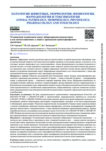
Эффективное лечение уролитиаза кошек во многом зависит от ранней диагностики заболевания, однако диагностический алгоритм этого вида патологии крайне ограничен в силу разных причин, в частности, он не учитывает наличие взаимосвязи в оси «почки-кишечник». Анализ функционального состояния гастроинтестинальной системы и состава микробиоты кишечника может повысить диагностический потенциал при уролитиазе, предсказать риск, улучшить прогноз и выбрать правильную стратегию лечения. Целью данной публикации является установление взаимосвязи между лабораторными показателями в оси «почки-кишечник» у кошек при мочекаменной болезни струвитного типа с признаками цистолитиаза.
Идентификаторы и классификаторы
Мочекаменная болезнь или уролитиаз — одна из наиболее распространенных патологий
мочевыделительной системы у кошек, эффективность лечения которой во многом зависит от ранней диагностики [1–5]. Однако диагностический алгоритм уролитиаза на ранних стадиях затруднен в силу выраженных компенсаторных механизмов организма при поражении почек, а наличие взаимосвязи в оси «почки-кишечник» практически не учитывается [6–12]. Изучением взаимосвязи между кишечником и урогенитальным трактом в последние годы много занимаются в гуманной медицине: доказано, что между микробиотой кишечника и функциональной активностью почек существует двунаправленная взаимосвязь, так как нарушение работы почек вызывает дисбактериоз кишечника, который, в свою очередь, ведет к развитию осложнений и прогрессированию заболевания вследствие развития эндотоксемии и ацидоза [13–23].
Список литературы
1. Filipska A, Bohdan B, Wieczorek PP, Hudz N. Chronic Kidney Disease and Dialysis Therapy: Incidence and Prevalence in the World. Pharmacia. 2021;68(2):463-470. https://doi.org/10.3897/pharmacia.68.e65501
2. Соболев В.Е. Нефрология и урология домашней кошки (Felis catus). Российский ветеринарный журнал. Мелкие домашние и дикие животные. 2011;1:40-42.
3. Knoll T, Schönthaler M, Neisius A. Urolithiasis. Der Urologe. 2019;58:1271. https://doi.org/10.1007/s00120-019-01047-1
4. Kirkali Z, Rasooly R, Star RA, Rodgers GP. Urinary Stone Disease: Progress, Status, and Needs. Urology. 2015;86(4):651-653. https://doi.org/10.1016/j.urology.2015.07.006
5. Bacârea A, Fekete GL, Grigorescu BL, Bacârea VC. Discrepancy in Results between Dipstick Urinalysis and Urine Sediment Microscopy. Experimental and Therapeutic Medicine. 2021;21(5):538. https://doi.org/10.3892/etm.2021.9971
6. Gottlieb M, Long B, Koyfman A. The Evaluation and Management of Urolithiasis in the ED: A Review of the Literature. The American Journal of Emergency Medicine. 2018;36(4): 699-706. https://doi.org/10.1016/j.ajem.2018.01.003.
7. Hernandez N, Song Y, Noble VE, Eisner BH. Predicting Ureteral Stones in Emergency Department Patients with Flank Pain: An External Validation of the STONE Score. World Journal of Urology. 2016;34:1443-1446. https://doi.org/10.1007/s00345-016-1760-3
8. Safaie A, Mirzadeh M, Aliniagerdroudbari E, Babaniamansour S, Baratloo A. A Clinical Prediction Rule for Uncomplicated Ureteral Stone: The STONE Score; A Prospective Observational Validation Cohort Study. Turkish Journal of Emergency medicine. 2019;19(3):91-95. https://doi.org/10.1016/j.tjem.2019.04.001
9. Kopecny L, Palm CA, Segev G, Larsen JA, Westropp JL. Urolithiasis in Cats: Evaluation of Trends in Urolith Composition and Risk Factors (2005-2018). Journal of Veterinary Internal Medicine. 2021;35(3):1397-1405. https://doi.org/10.1111/jvim.16121
10. Gambaro G, Croppi E, Bushinsky D, Jaeger P, Cupisti A, Ticinesi A, et al. The Risk of Chronic Kidney Disease Associated with Urolithiasis and its Urological Treatments: A Review. The Journal of Urololy. 2017;198(2): 268-273. https://doi.org/10.1016/j.juro.2016.12.135
11. Ватников Ю.А., Миколенко О.Н., Вилковыский И.Ф., Паршина В.И., Трошина Н.И. Динамика биохимических показателей сыворотки крови при мочекаменной болезни у кошек. Лечение. Ветеринария, зоотехния и биотехнология. 2016;(12):48-54.
12. Мелешков С.Ф. Динамика функциональных расстройств мочеиспускания и их клинико-моРоссийская Федерацияологические параллели при урологическом синдроме у кошек. Ветеринарная патология. 2008;(3):48-55.
13. Almannie RM, Alsufyani AK, Alturki AU, Almuhaideb M, Binsaleh S, Althunayan AM, et al. Neural Network Analysis of Crystalluria Content to Predict Urinary Stone Type. Research and Reports in Urology. 2021;13:867-876. https://doi.org/10.2147/rru.S322580
14. Abufaraj M, Al Karmi J, Yang L. Prevalence and Trends of Urolithiasis among Adults. Current Opinion in Urology. 2022;32(4):425-432. https://doi.org/10.1097/mou.0000000000000994
15. Kaul E, Hartmann K, Reese S, Dorsch R. Recurrence Rate and Long-Term Course of Cats with Feline Lower Urinary Tract Disease. Journal of Feline Medicine and Surgery. 2020;22(6):544-56. https://doi.org/10.1177/1098612X19862887
16. Gomes VDR, Ariza PC, Borges NC, Schulz Jr. J, Fioravanti MCS. Risk Factors Associated with Feline Urolithiasis. Veterinary Research Communications. 2018;42(1):87-94. https://doi.org/10.1007/s11259-018-9710-8
17. Ушакова Т.М. Роль гепаторенальной системы в развитии метаболических нарушений у кошек, больных трипельфосфатным уролитиазом. Известия Оренбургского государственного аграрного университета. 2019;6(80):199-202.
18. Ушакова Т.М., Дерезина Т.Н. Урологический и клинический статусы кошек под действием комплексной фармакокоррекции уролитиаза на фоне диетотерапии. Вестник Донского государственного аграрного университета. 2018;(3-1(29)):5-12.
19. Ушакова Т.М., Старикова Е.А., Дерезина Т.Н. Комплексный алгоритм фармакокоррекции расстройств гепаторенальной системы у кошек, больных уролитиазом. Ветеринарная патология. 2019;(4):28-38.
20. Cleroux A, Alexander K, Beauchamp G, Dunn M. Evaluation for Association between Urolithiasis and Chronic Kidney Disease in Cats. Journal of the American Veterinary Medical Association. 2017;250(7): 770-774. https://doi.org/10.2460/javma.250.7.770
21. Burggraaf ND, Westgeest DB, Corbee RJ. Analysis of 7866 Feline and Canine Uroliths Submitted between 2014 and 2020 in the Netherlands. Research in Veterinary Science. 2021;137:86-93. https://doi.org/10.1016/j.rvsc.2021.04.026
22. Hobby GP, Karaduta O, Dusio GF, Singh M, Zybailov BL, Arthur JM. Chronic Kidney Disease and the Gut Microbiome. American Journal of Physiology-Renal Physiology. 2019;316(6):F1211-F1217. https://doi.org/10.1152/ajprenal.00298.2018
23. Jacobson DK, Honap TP, Ozga AT, Meda N, Kagoné TS, Carabin, et al. Analysis of Global Human Gut Metagenomes Shows that Metabolic Resilience Potential for Short-Chain Fatty Acid Production is Strongly Influenced by Lifestyle. Scientific Reports. 2021;11:1724. https://doi.org/10.1038/s41598-021-81257-w
24. Mao L, Franke J. Symbiosis, Dysbiosis, and Rebiosis-The Value of Metaproteomics in Human Microbiome Monitoring. Proteomics. 2015;15:1142-1151. https://doi.org/10.1002/pmic.201400329
25. Li M, Wang B, Zhang M, Rantalainen M, Wang S, Zhou H, et al. Symbiotic Gut Microbes Modulate Human Metabolic Phenotypes. PNAS. 2008;105(6): 2117-2122. https://doi.org/10.1073/pnas.0712038105
26. Berg G, Rybakova D, Fischer D, Cernava T, Vergès MCC, Charles T, et al. Microbiome Definition Re-Visited: Old Concepts and New Challenges. Microbiome. 2020;8:103. https://doi.org/10.1186/s40168-020-00875-0
Выпуск
Другие статьи выпуска
Архивы государственных библиотек Португалии и Испании хранят несколько значимых произведений, созданных практикующими ветеринарными врачами XVII века и посвященных лечению лошадей, мулов и ослов. В этих трудах, которые представлены в отсканированном виде на официальных сайтах библиотек, описаны причины возникновения болезней животных, их клинические признаки и методы лечения. Одним из часто упоминающихся патологических состояний животных средневековой Испании являются раны. В статье приведен анализ некоторых из этих интереснейших научных источников с целью установления методик диагностики и лечения ран лошадей, а также для выявления исторического вектора развития ветеринарной медицины с учетом современного ее состояния в изучаемой области научно-практического знания.
Автомобильный инцидент является одной из самых распространенных причин травмирования собак: согласно зарубежной статистике, не менее 51 % от общего количества случаев травм собак. Основную группу риска составляют кобели в возрасте от 1 до 3 лет. В России отсутствуют исследования распространённости автомобильных травм у собак, которые позволили бы определить факторы риска, характер и тяжесть повреждений, сформировать рекомендации для владельцев и ветеринарных врачей. В настоящей работе предлагается ретроспективный анализ распространённости автомобильных травм у собак на основании данных сети ветеринарных клиник Ростовской области за 2018–2022 гг.
Массометрия и вычисление массовых коэффициентов внутренних органов лабораторных животных является неотъемлемым этапом при проведении токсикологических исследований. Однако при отсутствии корректных внутрилабораторных референтных интервалов, которые бы отражали нормальные значения для популяции животных исследовательского центра, анализ изменений по массовым коэффициентам в эксперименте может быть весьма затруднителен. В частности, речь идет о данных по массам органов и массовым коэффициентам органов карликовых свиней, которые в открытых источниках представлены недостаточно. Однако присутствует множество такого рода данных в литературе, касающейся промышленных пород свиней. Ввиду того, что карликовые свиньи широко используются при проведении доклинических исследований и при этом рассматриваются как одна из весовых категорий в систематике видов свиней, представляющая собой свинью со сниженной массой тела со схожим строением органов и систем организма, нами была поставлена цель — рассчитать референтные интервалы массовых коэффициентов внутренних органов, вычисленных относительно массы тела и головного мозга, а также абсолютных значений массы внутренних органов карликовых свиней, и провести их сравнение с данными из литературных источников по карликовым и промышленным породам свиней.
Для аэрозольной дезинфекции птицеводческих помещений применяется огромное количество дезинфектантов, большинство из которых рекомендованы для профилактической или заключительной дезинфекции в отсутствие животных. Тем не менее некоторые средства, имеющиеся в арсенале ветеринарных служб, имеют рекомендации по текущей дезинфекции и применяются в присутствии птицы, хотя далеко не все из них отвечают предъявляемым требованиям с точки зрения состава, целей и режимов их использования. В частности, интерес представляет дезинфектант на основе глутарового альдегида и четвертичных аммониевых соединений, уже рассматривавшийся в научных источниках: не до конца изученным остался вопрос влияния этого препарата на физиологический и зоотехнический статус птицы, находящейся в зоне обработки этим средством. Целью данного исследования является изучение физиологического статуса и продуктивных качеств цыплятбройлеров, подвергшихся непосредственному воздействию дезинфицирующего средства на основе глутарового альдегида и четвертичных аммониевых соединений в режиме аэрозольного распыления.
Необходимость моделирования оксидативного стресса в эксперименте воздействием переменного магнитного поля низкой частоты связана с постоянным увеличением электромагнитной нагрузки на теплокровный организм ввиду ежегодного ухудшения электромагнитного состояния внешней среды. Переменное магнитное поле низкой частоты запускает каскад биохимических реакций у лабораторных животных, изменяющих гомеостаз на фоне повышения интенсивности свободнорадикального (перекисного) окисления липидов биомембран. Препараты, содержащие янтарную кислоту, обладают антиоксидантным, антигипоксантным, актопротекторным и стресс-протективным действием, апробированным в различных модельных системах, однако отсутствие данных об эффективности янтарной кислоты в условиях воздействия переменного магнитного поля стало основанием для проведения настоящего эксперимента. Цель данного исследования — определение защитных эффектов янтарной кислоты при воздействии переменного магнитного поля низкой частоты на лабораторных крыс.
Класс Cestoda подразделяется на два подкласса: Cestodaria — нечленистых ленточных червей и Eucestoda — настоящих цестод. У хищных млекопитающих паразитируют представители отрядов Pseudophyllidea и Cyclophyllidae, входящие в подкласс настоящих цестод. При этом у рукокрылых паразитируют только представители последнего отряда. Данные о видовом составе и распространении цестод в Ростовской области до сих пор не были опубликованы. Ранее был составлен список видов летучих мышей, обитающих в Ростовской области. В настоящей статье авторы рассматривают фауну цестод, паразитирующих в рукокрылых исследуемого региона. Целью данного обзора является анализ современного состояния изученности видового состава гельминтов, паразитирующих у рукокрылых, обитающих на территории Ростовской области.
Статистика статьи
Статистика просмотров за 2025 - 2026 год.
Издательство
- Издательство
- ДГТУ
- Регион
- Россия, Ростов-на-Дону
- Почтовый адрес
- 344003, ЮФО, Ростовская область, г. Ростов-на-Дону, пл. Гагарина, 1
- Юр. адрес
- 344003, Ростовская обл, г Ростов-на-Дону, пл Гагарина, зд 1
- ФИО
- Месхи Бесарион Чохоевич (РЕКТОР)
- E-mail адрес
- reception@donstu.ru
- Контактный телефон
- +8 (800) 1001930
- Сайт
- https://donstu.ru


